
Page Index:
The Ultimate Guide to HbA1c Testing
This page and all its content are intended solely for educational purposes, to provide insight into the subject of HbA1c Testing. Sky Health Wellness Clinic does not promote or endorse any specific treatments, medications, or pharmaceutical brands mentioned herein. Any external links provided are for informational context only and do not imply endorsement.
While this page provides information on the HbA1c Testing and its potential health applications, it is not a substitute for your primary care provider's medical advice. Always consult your primary care provider or a licensed healthcare professional before making decisions about your health, including any treatment options or medication use. For more information, please visit our Medical Disclaimer Page.
If you have questions, identify any inaccuracies, or would like to suggest an update, please reach out to us through our website’s contact form. Our team is committed to providing accurate, up-to-date, and helpful information to support your wellness journey. For the best user experience, try using the desktop view.
This content has been reviewed and approved by our team of medical providers.
This page was last updated: 05/13/2026

HbA1c Testing FAQ

The Physiological Basis of the HbA1c Test
The Hemoglobin A1c (HbA1c) test, also known as glycated hemoglobin or glycosylated hemoglobin, is the primary clinical biomarker used to determine the average blood glucose levels of an individual over a period of approximately two to three months. Unlike traditional capillary or venous glucose measurements—which provide a momentary assessment of glucose concentration at the specific time of the blood draw—the HbA1c provides a retrospective analysis of glycemic control.
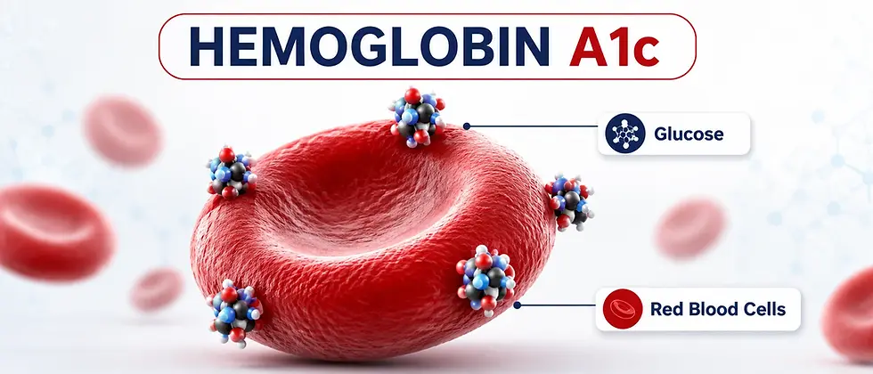
The biological utility of this test stems from the lifespan of the erythrocyte (red blood cell). In a healthy human adult, red blood cells circulate for an average of 100 to 120 days before being sequestered and recycled by the spleen. Throughout this circulation period, hemoglobin proteins within the red blood cells are exposed to plasma glucose. Through a spontaneous chemical process, glucose molecules adhere to the hemoglobin, creating a record of the glucose environment that persists until the cell is destroyed.
Diagnostic Thresholds and Clinical Utility
The HbA1c test is the gold standard for diagnosing metabolic disorders and monitoring treatment efficacy in diabetic populations. According to established international guidelines, the following ranges define metabolic status:
-
Normal: Below 5.7%.
-
Prediabetes: 5.7% to 6.4%.
-
Diabetes: 6.5% or higher.
For patients already diagnosed with diabetes, the A1c is used to assess the risk of long-term microvascular complications. Sustained elevations in A1c are directly correlated with an increased risk of retinopathy (eye damage), nephropathy (kidney disease), and neuropathy (nerve damage). Conversely, every 1% reduction in HbA1c levels has been shown to significantly decrease the risk of these complications.
The "Video" vs. "Snapshot" Comparison
In clinical terms, a fasting plasma glucose (FPG) test is a "snapshot." It can be influenced by acute factors such as stress, recent exercise, illness, or a high-carbohydrate meal consumed the night before the test. The HbA1c is a "video." Because the bond between glucose and hemoglobin is stable and irreversible, the test is unaffected by short-term fluctuations. This makes it an invaluable tool for identifying "silent" hyperglycemia in patients who may have normal fasting levels but experience significant glucose spikes after meals or during the night.

The Chemical Compound & Biological Mechanism
(Glycation Science)
To understand the HbA1c test at a molecular level, one must examine the protein chemistry of hemoglobin and the spontaneous reaction known as the Maillard reaction.
The Structure of Hemoglobin A
Hemoglobin is a metalloprotein found in red blood cells that is responsible for the transport of oxygen. In adults, the most common form is Hemoglobin A (HbA), which accounts for approximately 95%–97% of total hemoglobin. HbA is a tetramer consisting of two alpha (α) polypeptide chains and two beta (β) polypeptide chains.
The Chemistry of Non-Enzymatic Glycation
The formation of HbA1c is a non-enzymatic process, meaning it does not require a biological catalyst (enzyme) to occur. Instead, it is a concentration-dependent chemical reaction. The higher the concentration of glucose in the plasma, the higher the percentage of hemoglobin that becomes glycated.
This reaction occurs in three distinct chemical stages:
-
Formation of the Schiff Base (Pre-A1c): Glucose in the blood exists in an equilibrium between a ring structure and an open-chain aldehyde form. The aldehyde group of the glucose molecule reacts with the free amino group (specifically the N-terminal valine) of the hemoglobin beta chain. This initial reaction is rapid and reversible, resulting in an unstable intermediate known as an aldimine, or "Schiff base".
-
The Amadori Rearrangement: Over a period of days, the unstable Schiff base undergoes a slow, spontaneous structural change known as an Amadori rearrangement. During this process, the aldimine is converted into a more stable ketoamine.
-
The Final Stable Product: Once the Amadori rearrangement is complete, the glucose is "locked" onto the hemoglobin protein. This final, stable ketoamine is what we measure as HbA1c. Unlike the Schiff base, this compound will remain attached to the hemoglobin for the entire 120-day lifespan of the red blood cell.
Glycation vs. Glycosylation
It is scientifically critical to distinguish between glycation and glycosylation.
-
Glycosylation is a controlled, enzyme-mediated process where a carbohydrate is attached to a protein or lipid for a specific biological function (such as cell signaling).
-
Glycation (the process of forming HbA1c) is an uncontrolled, accidental reaction that occurs simply because of the presence of sugar in a high-oxygen environment.
Advanced Glycation End-Products (AGEs)
While HbA1c is the most commonly measured glycated protein, glycation can happen to almost any protein in the body. If blood sugar remains chronically high, these glycated proteins can undergo further chemical changes, eventually becoming Advanced Glycation End-products (AGEs).
AGEs are pathogenic; they cross-link with other proteins, making tissues stiff and less functional. They are a primary driver of the aging process and the structural damage seen in the blood vessels and organs of diabetic patients. HbA1c serves as the clinical "proxy" for the level of AGE-related damage occurring throughout the entire body.
The Role of the N-Terminal Valine
While glucose can attach to various points on the hemoglobin molecule (such as lysine residues), the HbA1c test specifically measures the glycation of the N-terminal valine residue of the beta chain. This specific site is the most reliable indicator of long-term glucose exposure and is the focus of all standardized laboratory assays.

The History and Origin of The HbA1c Test:
From Discovery to Diagnostic Gold Standard
The evolution of the Hemoglobin A1c test from a molecular curiosity to the definitive metric for metabolic health represents one of the most significant advancements in 20th-century medicine. Before the 1970s, the clinical management of blood sugar was largely reactive, relying on erratic "snapshots" that failed to capture the true trajectory of a patient's glycemic state.
The Era of Qualitative Testing
Before the mid-1900s, clinicians relied primarily on urine glucose testing. This method was pioneered using reagents like Benedict’s solution, which changed color in the presence of sugar when heated. While revolutionary for the time, urine testing was profoundly limited by the "renal threshold"—glucose typically does not appear in the urine until blood concentrations exceed 180 mg/dL. Consequently, patients could remain in a state of chronic, moderate hyperglycemia for years without a single positive urine test, leading to undetected systemic damage.
Discovery of Hemoglobin Heterogeneity (1950s)
The scientific foundation for the HbA1c test was laid in 1955 when researchers Kunkel and Wallenius utilized starch-gel electrophoresis to study human hemoglobin. They discovered that adult hemoglobin was not a single, uniform protein but was composed of several "minor" fractions. These fractions were labeled HbA1a, HbA1b, and HbA1c, depending on their migration speed during electrophoresis. At the time of discovery, the chemical reason for these variations remained unknown; they were viewed as biochemical anomalies rather than diagnostic tools.
The Rahbar Breakthrough (1968–1969)
The pivotal link between these hemoglobin fractions and glucose metabolism was discovered by Dr. Samuel Rahbar in Tehran, Iran. While screening 1,200 individuals for hemoglobin variants, Rahbar observed an abnormally high "fast-moving" hemoglobin band in two diabetic patients. Further investigation of 47 diabetic subjects confirmed that this specific fraction—HbA1c—was consistently elevated to two to three times the level found in non-diabetic individuals. Rahbar’s work proved that HbA1c was not a genetic defect but a physiological consequence of high blood sugar.
Clinical Validation and Cerami's Contribution (1976)
In 1976, Dr. Anthony Cerami and his colleagues at Rockefeller University established the correlation between HbA1c and long-term glucose averages. They demonstrated that because glycation is slow and irreversible, the A1c level serves as a "molecular memory" of the previous 120 days. This discovery allowed physicians, for the first time, to distinguish between a patient who had a single "good" fasting blood sugar on the day of their appointment and a patient who maintained stable control consistently.
The Path to Standardization (1990s–2026)
By the early 1990s, HbA1c testing was widespread, but laboratory results lacked uniformity. A patient could receive a 7% result at one lab and a 9% result at another, leading to dangerous inconsistencies in treatment. This led to the creation of the National Glycohemoglobin Standardization Program (NGSP) in 1996, which mandated that all laboratory assays be traceable to the reference methods used in major clinical trials. Today, in 2026, international harmonization ensures that an A1c result is universally interpreted, regardless of where the test is performed.

HbA1c Test Clinical Trials and Landmark Studies:
The Evidence That Changed Medicine
The transition of HbA1c from a "monitoring tool" to a "diagnostic standard" was driven by massive, multi-decade clinical trials. These studies proved that HbA1c levels were not just numbers, but direct predictors of life-altering complications.
The DCCT: Proving the Link in Type 1 Diabetes (1983–1993)
The Diabetes Control and Complications Trial (DCCT) is perhaps the most influential study in the history of endocrinology. It followed 1,441 participants with Type 1 Diabetes over a decade to determine if "intensive control" (lowering A1c as much as possible) actually prevented damage.
-
The Result: The study found that intensive control, which maintained an average A1c of approximately 7%, resulted in a 76% reduction in the risk of retinopathy (eye disease) compared to the standard care group (average A1c of 9%).
-
Impact: It established the HbA1c level of 7% as the universal target for preventing microvascular damage.
The UKPDS: Proving the Link in Type 2 Diabetes (1977–1997)
While the DCCT focused on Type 1, the United Kingdom Prospective Diabetes Study (UKPDS) spent 20 years studying 5,102 patients with Type 2 Diabetes.
-
Key Findings: For every 1% reduction in HbA1c, the risk of microvascular complications (kidney and eye damage) dropped by 37%, and the risk of diabetes-related death dropped by 21%.
-
Legacy Effect: Even more remarkably, follow-up studies showed that patients who maintained low A1c levels early in their diagnosis continued to have lower risks of heart attack and death decades later, even if their control slipped later on. This phenomenon is known as "Metabolic Memory".
The Intensive Control Debate: ACCORD, ADVANCE, and VADT
In the late 2000s, three major trials (ACCORD, ADVANCE, and VADT) investigated whether lowering A1c even further—below 6.0%—was beneficial.
-
Unexpected Risks: The ACCORD trial was famously halted early because the group with the lowest A1c targets (intensive group) actually showed a higher rate of mortality.
-
Clinical Nuance: These trials taught the medical community that "lower is not always better" for every patient, especially older adults with long-standing heart disease who are at high risk for hypoglycemia (dangerously low blood sugar).
Current Consensus on The HbA1c Test (2026)
These trials have led to the personalized medicine approach we use in 2026. While the general target remains below 7%, clinicians now adjust targets based on a patient’s age, life expectancy, and other health conditions, using the historical data from these landmark trials as a roadmap.

How The HbA1c Test is Made:
Laboratory Assays and Manufacturing Technology
In 2026, the technology used to manufacture HbA1c assays has reached a level of precision that allows for near-instantaneous results with minimal margin for error. The production of these tests involves complex biotechnology and chemical engineering.
The Chemical Components of an A1c Assay
An HbA1c test kit typically consists of several highly specific chemical reagents:
-
Lysis Buffers: These are detergents designed to break open the red blood cells (hemolysis), releasing the hemoglobin into the solution so it can be measured.
-
Specific Antibodies (Immunoassays): Many modern tests use monoclonal antibodies that are engineered to recognize and bind only to the glycated N-terminal valine of the hemoglobin beta chain.
-
Enzymatic Reagents: Some tests utilize enzymes like fructosyl peptide oxidase, which react with the glycated amino acids to produce a color change that can be measured by a machine.
Manufacturing Methods: The Three Primary Technologies
There are three main ways the HbA1c test is manufactured and executed in 2026:
-
Ion-Exchange High-Performance Liquid Chromatography (HPLC): This is considered the "reference method." It works by separating the hemoglobin fractions based on their electrical charge. Because glycated hemoglobin has a different charge than non-glycated hemoglobin, it moves through the liquid column at a different speed.
-
Immunoassays: These tests use the antibodies mentioned above. When the antibody binds to the HbA1c, it forms a "clump" (agglutination) that blocks light. The machine measures how much light is blocked to calculate the percentage of A1c.
-
Boronate Affinity Chromatography: This method uses a chemical called boronate that has a natural "stickiness" for glucose. It pulls all glycated hemoglobin out of the sample, regardless of where the glucose is attached, providing a very stable measurement that is less affected by common blood variants.

Understanding Your HbA1c Test Results:
Percentages, eAG, and Target Goals
Interpreting an HbA1c result requires a transition from understanding simple percentages to understanding how those percentages translate into daily physiological experiences. Because the HbA1c is a weighted average—with approximately 50% of the value determined by the most recent 30 days of glucose exposure—clinicians use several different metrics to explain these results to patients.
The Diagnostic Categories (2026 Standards)
The medical community maintains strict consensus on the thresholds for metabolic health. These numbers are utilized globally to categorize a patient’s risk profile:
-
Normal (Healthy Range): Below 5.7% A result in this range indicates that the body is efficiently managing glucose through insulin sensitivity and appropriate pancreatic response. Glycation of hemoglobin is occurring at a baseline rate consistent with non-pathological aging.
-
Prediabetes (Increased Risk): 5.7% to 6.4% This range is a critical clinical indicator of "impaired glucose tolerance" or "impaired fasting glucose." It signifies that the body’s metabolic machinery is under stress. At this level, the risk of progressing to full-blown Type 2 Diabetes is high, but the condition is often reversible through intensive lifestyle and nutritional intervention.
-
Diabetes Mellitus: 6.5% or Higher A result of 6.5% or higher on two separate tests (to rule out laboratory error or acute interference) is the definitive diagnostic criterion for diabetes. At this level, the concentration of glucose in the blood is high enough to cause progressive microvascular damage over time.
The Estimated Average Glucose (eAG)
One of the most significant barriers to patient understanding was the "percentage" unit itself. Patients are accustomed to seeing blood sugar numbers in mg/dL (milligrams per deciliter) on their home glucometers, making a "7.0%" result feel abstract. To bridge this gap, the ADAG (A1c-Derived Average Glucose) study established a mathematical formula to convert the A1c percentage into an Estimated Average Glucose (eAG).
The standardized formula used in 2026 for this conversion is:
28.7 X HbA1c - 46.7 = eAG (in mg/dL)
eAG Conversion Table:
HbA1c (%) | eAG (mg/dL) | eAG (mmol/L) |
|---|---|---|
5 | 97 | 5.4 |
5.5 | 111 | 6.2 |
6 | 126 | 7 |
6.5 | 140 | 7.8 |
7 | 154 | 8.6 |
7.5 | 169 | 9.4 |
8 | 183 | 10.2 |
9 | 212 | 11.8 |
10 | 240 | 13.4 |
The Estimated Average Glucose (eAG)
While a "Normal" A1c is below 5.7%, the "Target" for a patient already living with diabetes is not always to reach that number. Overtreatment can lead to hypoglycemia (dangerously low blood sugar), which can be acutely fatal or cause cognitive decline.
Current 2026 clinical guidelines for A1c targets include:
-
Healthy Adults: A general goal of < 7.0% is often recommended to prevent long-term complications while minimizing the risk of "hypo" events.
-
Elderly or Frail Patients: Targets may be relaxed to 7.5% or 8.0%. In these populations, the immediate danger of a fall or seizure caused by low blood sugar outweighs the long-term risk of microvascular damage.
-
Pregnant Patients: Due to the high stakes of fetal development, targets are much tighter, often seeking an A1c of < 6.0% or < 6.5%.
-
Pediatric Patients: Goals are typically < 7.0%, with an emphasis on using technology like Continuous Glucose Monitors (CGMs) to avoid glycemic variability.
The Limitation: Glycemic Variability
It is vital to note that two patients can have the exact same A1c of 7.0% but very different health outcomes.
-
Patient A may have "stable" blood sugar that rarely moves outside the 140–160 mg/dL range.
-
Patient B may have "volatile" blood sugar that swings from 40 mg/dL (dangerously low) to 300 mg/dL (dangerously high).
Because A1c is an average, it "masks" these dangerous swings. In 2026, clinicians increasingly use Time in Range (TIR)—the percentage of the day spent between 70 and 180 mg/dL—as a necessary companion to the HbA1c test.

Factors Affecting the Accuracy of HbA1c Test:
The Biological "Noise"
The HbA1c test is highly reliable, but it is not infallible. Because the test relies on the mathematical assumption that every patient's red blood cells live for exactly 120 days, any condition that alters red blood cell (RBC) lifespan or hemoglobin structure will produce a "false" A1c reading.
Red Blood Cell Lifespan Alterations
If red blood cells do not live long enough, they have less time to "collect" sugar, leading to a falsely low A1c. Conversely, if they live too long, they collect more sugar than average, leading to a falsely high A1c.
-
Falsely Low A1c Causes:
-
Hemolytic Anemia: RBCs are destroyed prematurely.
-
Recent Blood Loss or Transfusion: The presence of new, young RBCs lowers the overall glycation percentage.
-
Erythropoietin (EPO) Therapy: Common in kidney patients, this stimulates the rapid production of new RBCs.
-
-
Falsely High A1c Causes:
-
Iron, B12, or Folate Deficiency Anemia: In these states, RBC production slows down, meaning the existing cells stay in circulation longer than usual.
-
Asplenia: Patients without a spleen (which recycles old RBCs) may have older-than-average cell populations.
-
Hemoglobin Variants (Hemoglobinopathies)
Approximately 7% of the global population carries a hemoglobin variant, such as HbS (Sickle Cell), HbC, or HbE.
-
In the past, these variants interfered with the chemical "migration" in HPLC tests, making the results unreadable.
-
In 2026, modern Immunoassays and Boronate Affinity tests are generally "variant-blind," meaning they can provide accurate results even in patients with these traits. However, patients with Sickle Cell Disease (not just the trait) still face accuracy issues due to significantly shortened RBC lifespans.
Pregnancy and Hormonal Shifts
During pregnancy, RBC turnover increases, and blood volume expands. This typically results in a physiologically lower A1c. A "normal" A1c in a non-pregnant woman might actually indicate gestational diabetes in a pregnant woman.
Chronic Kidney Disease (CKD)
Kidney failure is a "perfect storm" for A1c inaccuracies. CKD patients often suffer from shortened RBC lifespans, are frequently treated with EPO, and may have carbamylated hemoglobin—a structural change caused by urea that can "mimic" glycated hemoglobin in certain older lab tests.
Chemical and Supplement Interference
-
High-Dose Vitamin C or E: Some studies suggest that very high doses of antioxidants may inhibit the glycation process itself, leading to a lower A1c that does not reflect actual blood sugar levels.
-
Alcoholism and Liver Disease: High intake of alcohol can lead to the formation of acetaldehyde-hemoglobin adducts, which may interfere with certain assay methods.

HbA1c and Longevity:
Beyond the Diabetic Population
In recent years, the clinical utility of the HbA1c test has expanded beyond the diagnosis and management of diabetes to serve as a predictive marker for longevity and all-cause mortality in the general population. While the medical community historically focused on the "6.5% threshold" for disease, emerging research in 2026 suggests that the relationship between glycation and health outcomes exists on a continuous spectrum, even within the "normal" range.
The Specter of "Normal-High" HbA1c
Research indicates that individuals with HbA1c levels at the high end of the normal range (e.g., 5.4% to 5.6%) may face higher risks of cardiovascular disease and cognitive decline compared to those at the lower end (e.g., 4.8% to 5.2%). This is often attributed to the cumulative effect of postprandial (after-meal) glucose spikes that do not yet reach the diagnostic criteria for prediabetes but still contribute to systemic oxidative stress and endothelial dysfunction.
-
Cardiovascular Risk: Large-scale observational studies have demonstrated that for every 1% increase in HbA1c within the non-diabetic range, there is a measurable increase in the risk of coronary heart disease.
-
Neurodegeneration: Chronic exposure to even slightly elevated glucose levels is associated with brain atrophy and an increased risk of developing Alzheimer’s disease and other forms of dementia, a phenomenon sometimes referred to in clinical circles as "Type 3 Diabetes".
Glycation and the Biological Clock
At the heart of the link between HbA1c and longevity is the process of glycation. As discussed in Section 2, glycation leads to the formation of Advanced Glycation End-products (AGEs). In the context of longevity, AGEs act as "metabolic glue," cross-linking with long-lived proteins like collagen and elastin.
This cross-linking results in:
-
Arterial Stiffness: Reduced elasticity in the blood vessels, leading to hypertension.
-
Skin Aging: The breakdown of dermal collagen, manifesting as wrinkles and loss of structural integrity.
-
Organ Decline: Progressive stiffening of the extracellular matrix in the heart, kidneys, and lungs.
The U-Shaped Curve of Mortality
Interestingly, longevity data often show a "U-shaped" relationship between HbA1c and mortality. While high A1c levels (above 6.5%) are associated with clear risks, extremely low A1c levels (below 4.0% to 4.5%) in non-diabetic individuals have also been linked to increased mortality in some studies. These very low levels may not be caused by "superior" glucose control but rather by underlying conditions such as occult malignancy, chronic liver disease, or certain types of anemia that falsely lower the A1c reading. Thus, in 2026, the "sweet spot" for metabolic longevity is generally considered to be between 4.8% and 5.3%.
Molecular Variations and the Genetics of Hemoglobin
While the standardized HbA1c test assumes a baseline of Hemoglobin A (HbA), human genetics introduces a wide array of hemoglobin variants that can fundamentally alter the molecular structure of the protein and its interaction with glucose. In 2026, understanding these genetic nuances is critical for personalized metabolic medicine, as a "standard" test may provide misleading data for millions of individuals worldwide.
The Diversity of Hemoglobin Types
Hemoglobin is not a static protein across the human population. There are several naturally occurring forms:
-
Hemoglobin A (HbA): The "normal" adult hemoglobin, comprising two alpha and two beta chains. This is the primary target of glycation measured in the HbA1c test.
-
Hemoglobin F (Fetal Hemoglobin): The primary hemoglobin in fetuses and newborns. It has a much higher affinity for oxygen and typically disappears within the first six months of life. However, some adults have "Hereditary Persistence of Fetal Hemoglobin" (HPFH). Because HbF does not glycate at the same rate as HbA, its presence can cause a falsely low HbA1c reading.
-
Hemoglobin S (Sickle Cell): Caused by a single amino acid substitution in the beta chain. Individuals with Sickle Cell Trait (one gene) or Disease (two genes) have red blood cells that are structurally fragile.
How Genetic Variants Interfere with Measurement
Genetic variations in hemoglobin interfere with the HbA1c test through two distinct mechanisms: chemical interference and biological interference.
-
Chemical Interference: In older laboratory methods like Ion-Exchange HPLC, variants like HbS, HbC, or HbE would "migrate" at the same speed as glycated HbA. This led to overlapping peaks on the laboratory chromatogram, making it impossible for the machine to distinguish between the variant and the sugar-coated protein.
-
Biological Interference: This is more complex and occurs even with the most advanced "variant-blind" assays. If a genetic variant causes red blood cells to live for 90 days instead of 120 days, the HbA1c will be falsely low because the cells simply haven't been in the bloodstream long enough to accumulate glucose.
Ethnic Variations in Baseline A1c
One of the most researched topics in 2026 is why certain ethnic groups—specifically African American, Hispanic, and Asian populations—often show higher baseline HbA1c levels than Caucasians, even when their average blood sugar (as measured by continuous glucose monitors) is identical.
-
Glycation Gap: Some researchers propose a "glycation gap," suggesting that intracellular glucose transport or the rate of the Amadori rearrangement (see Section 2) may be genetically programmed to happen faster in some individuals.
-
Red Cell Survival: Small differences in the average survival time of red blood cells across different populations can lead to a 0.1% to 0.4% difference in HbA1c that is not related to diet or exercise.
Addressing Hemoglobinopathies in the Lab
In 2026, most clinical laboratories use boronate affinity chromatography or specialized immunoassays to bypass these genetic hurdles. Boronate affinity is particularly effective because it binds to the glucose molecule itself, rather than the specific amino acid chain of the hemoglobin. This allows for an accurate measurement even if the underlying hemoglobin protein is structurally "abnormal".

The Future of Glycemic Monitoring: HbA1c vs. Alternatives
As we move further into 2026, the medical community is recognizing that while the HbA1c test is the gold standard, it is not the only standard. For patients where the A1c is unreliable (such as those with advanced kidney disease or severe anemia), several alternative markers are used to provide a complete metabolic picture.
Glycated Albumin and Fructosamine
Commonly known by the brand name GlycoMark, 1,5-AG is a specialized test that measures glucose spikes.
When to Use It: This is ideal for pregnant women with gestational diabetes, where rapid changes in glucose control need to be monitored more frequently than every three months. It is also used when starting a new medication to see if it’s working within weeks rather than months.
1,5-Anhydroglucitol (1,5-AG)
Commonly known by the brand name GlycoMark, 1,5-AG is a specialized test that measures glucose spikes.
-
The Mechanism: 1,5-AG is a naturally occurring sugar that the kidneys usually reabsorb. When blood sugar spikes above 180 mg/dL, the excess glucose blocks the reabsorption of 1,5-AG, causing levels in the blood to drop.
-
The Value: A patient can have a "perfect" A1c of 6.0% but have dangerously high spikes after meals. The 1,5-AG test will reveal these spikes even when the A1c average hides them.
The Shift to "Time in Range" (TIR)
With the widespread adoption of Continuous Glucose Monitors (CGMs), the medical community is shifting focus from "averages" to "consistency".
-
TIR Definition: The percentage of time a patient spends between 70 mg/dL and 180 mg/dL.
-
Predictive Power: 2026 studies have shown that Time in Range is as strongly correlated with kidney and eye complications as HbA1c. A patient with a 70% TIR is generally considered to be in excellent control, regardless of their A1c.
The Glycemic Management Indicator (GMI)
As mentioned in the FAQ, the GMI is a calculation that translates CGM data into an A1c-like number. In the future, many researchers believe the "lab-drawn" A1c may be replaced by the GMI for patients who wear sensors 24/7, as it provides a more personalized reflection of glucose exposure without the biological "noise" of red blood cell turnover.

Pediatric and Geriatric Specificities:
The Lifespan of Glycemic Control
As we move through 2026, clinical consensus has solidified around the idea that the HbA1c test cannot be interpreted through a "one-size-fits-all" lens across different age groups. The physiological environment of a growing child is fundamentally different from that of a senescing adult, and these differences necessitate distinct diagnostic thresholds and management strategies.
The Pediatric Population: Growth, Puberty, and Volatility
Managing and measuring glucose in children and adolescents presents unique challenges that directly influence HbA1c readings. In pediatric patients, especially those with Type 1 Diabetes, the goal is to balance strict glycemic control with the nutritional requirements of growth.
-
The Puberty Effect: During puberty, the body experiences a significant surge in growth hormones and sex steroids, both of which induce a temporary state of insulin resistance. This often leads to a "physiological climb" in HbA1c levels that is not necessarily due to poor dietary choices but rather to the hormonal milieu of adolescence.
-
Neurocognitive Development: In very young children (under age 5), the brain is highly sensitive to hypoglycemia (low blood sugar). Because the brain relies exclusively on glucose for energy, frequent "lows" can lead to developmental delays or cognitive impairment. Consequently, pediatric A1c targets in 2026 are often slightly more relaxed (typically around < 7.0%) than in non-pregnant adults to provide a safety buffer against severe hypoglycemia.
-
Glycemic Variability: Children often exhibit higher glycemic variability due to erratic activity levels and growth spurts. As discussed in Section 6, the A1c average can mask these dangerous swings, making the use of Continuous Glucose Monitors (CGMs) essential in this demographic.
The Geriatric Population: Senescence and Safety
In older adults (ages 65+), the interpretation of the HbA1c test shifts from "prevention of 30-year complications" to "preservation of current quality of life".
-
The Problem of Frailty: For a 20-year-old, an A1c of 8.5% is a significant risk for blindness in 20 years. For an 85-year-old, the primary risk is not blindness in two decades, but a fall or hip fracture tomorrow caused by a hypoglycemia-induced dizzy spell.
-
Modified Targets: In 2026, geriatric guidelines emphasize "de-intensification" of therapy. For older adults with multiple chronic conditions or cognitive impairment (such as dementia), an HbA1c target of 7.5% to 8.5% is often considered clinically superior to a strict < 7.0% target.
-
Altered RBC Dynamics: As humans age, the bone marrow’s efficiency in producing red blood cells may decline, and the prevalence of subclinical anemias increases. As discussed in Section 7, these changes can lead to falsely elevated or depressed A1c readings, requiring clinicians to verify A1c results with fructosamine or CGM data.

Lifestyle, Nutrition, and Dietary Influences on Glycation
While the HbA1c test is a biological measurement, it is profoundly influenced by external inputs. In 2026, the science of "glycemic load" and "oxidative stress" has provided a clearer picture of how specific lifestyle choices accelerate or decelerate the glycation of hemoglobin.
Nutritional Composition and the Glycemic Index
The rate at which hemoglobin becomes glycated is a direct function of the "area under the curve" (AUC) of daily blood glucose levels.
-
High Glycemic Index (GI) Foods: Foods that cause a rapid spike in blood sugar (refined sugars, white flours) provide a sudden, high concentration of glucose molecules to the bloodstream. This increases the probability of "Schiff base" formation (the first step of glycation).
-
Fiber and Protein "Buffering": Consuming fiber and protein alongside carbohydrates slows gastric emptying. This results in a "flatter" glucose curve, reducing the peak concentration of glucose and thus lowering the total glycation rate over time.
Exercise and "Non-Insulin Mediated" Glucose Uptake
Physical activity is perhaps the most potent tool for lowering HbA1c without pharmaceutical intervention. Exercise influences glycation through two primary mechanisms:
-
GLUT4 Translocation: Muscle contraction causes glucose transporter proteins (GLUT4) to move to the cell surface, allowing the muscles to "sop up" glucose from the blood without requiring extra insulin.
-
Increased Mitochondrial Density: Long-term aerobic and resistance training increases the number and efficiency of mitochondria, allowing the body to burn glucose more effectively as fuel rather than letting it circulate and glycate proteins.
The Role of Antioxidants and "Anti-Glycation" Nutrients
Emerging research in 2026 has investigated whether certain nutrients can inhibit the Amadori rearrangement (the second step of glycation). While not a replacement for glucose control, several compounds have shown promise in laboratory settings:
-
Benfotiamine: A fat-soluble version of Vitamin B1 that may help divert glucose into safer metabolic pathways, reducing the formation of AGEs.
-
Carnosine: A dipeptide found in muscle and brain tissue that may act as a "sacrificial" target for glycation, binding to glucose molecules before they can reach the hemoglobin.
-
Polyphenols: Found in green tea and berries, these compounds reduce the oxidative stress that typically accelerates the damage caused by glycated proteins.

HbA1c in Non-Diabetic Chronic Conditions
A major area of expansion in 2026 is the use of the HbA1c test to monitor health in patients who do not have diabetes but suffer from conditions characterized by insulin resistance or chronic inflammation.
Polycystic Ovary Syndrome (PCOS)
PCOS is a complex endocrine disorder where insulin resistance is a central feature. Even if a woman with PCOS has "normal" fasting glucose, her HbA1c may sit at the higher end of the normal range (e.g., 5.4%–5.6%). In these cases, the A1c is used as a tracker for the efficacy of metformin or inositol treatments and as an early warning system for the development of metabolic syndrome.
Sleep Apnea and Circadian Disruption
Obstructive Sleep Apnea (OSA) causes repeated bouts of intermittent hypoxia (low oxygen) and spikes in cortisol during the night. These cortisol spikes trigger the liver to release glucose while the patient is sleeping. Consequently, patients with untreated sleep apnea often have an unexpectedly high HbA1c relative to their daytime diet. In 2026, a "falling" HbA1c is often used as an objective marker that a patient’s CPAP therapy is working effectively.
Chronic Inflammation and Glucocorticoids
Conditions like rheumatoid arthritis or lupus often require long-term treatment with glucocorticoids (steroids like prednisone). Steroids are notorious for causing "steroid-induced hyperglycemia". For these patients, the HbA1c is a vital safety check to ensure that the treatment for their inflammation is not inadvertently causing permanent metabolic damage.

Psychosocial Impacts: "A1c Distress" and the Human Element
The HbA1c test is unique among medical tests because of the significant psychological weight it carries for patients. In 2026, clinicians are trained to recognize and mitigate "A1c-related distress".
The "Grade" Mentality
Because the A1c is presented as a single number every three months, many patients view it as a "grade" on their lifestyle or their "compliance". A "bad" A1c (e.g., rising from 7.2% to 7.8%) can lead to feelings of shame, failure, and "diabetes burnout," where the patient becomes so overwhelmed that they stop managing their condition altogether.
The Clinical Shift: From Judgment to Data
In 2026, the language surrounding the test has shifted. Rather than asking "Why is your A1c so high?", providers are encouraged to view the number as a neutral data point that reflects the intersection of biology, environment, and medication.
-
The Limitation of Control: Patients are taught that they only have "influence" over their A1c, not total "control," as factors like illness, stress, and genetics (Section 10) play a massive role.
-
The 2026 Focus: Success is increasingly defined by improvement and Time in Range rather than achieving a "perfect" 5.0%.